Lower Back Organ Anatomy Diagram : Kidney pain vs. Right, Left and Central Abdominal Pain - La vaca cega. Bones and cartilages of the external nose. It extends from the fourth to the sixth vertebral levels. We'll go over its different openings and functions before exploring the conditions that can affect the diaphragm. Nov 28, 2016 · anatomy of the larynx. Dec 06, 2017 · the primary cartilages in the human nose include the lateral, septal, and the greater and lesser alar cartilages 11.
The right and left sides of the heart are further divided into two top chambers called the atria (also termed the right and left atrium ), which receive blood and then pump it into the two bottom. Bones and cartilages of the external nose. The larynx, commonly called the voice box or glottis, is the passageway for air between the pharynx above and the trachea below. It extends from the fourth to the sixth vertebral levels. On the left is an anatomy diagram of the internal organs of a female elephant.
BACK MUSCLES ANATOMY | JACKED FOREVER from 2.bp.blogspot.com Ingested food exits the small intestine and enters the cecum. Figure 11.32 muscles of the lower leg the muscles of the anterior compartment of the lower leg are generally responsible for dorsiflexion, and the muscles of the posterior compartment of the lower leg are generally responsible for plantar flexion. The right and left sides of the heart are further divided into two top chambers called the atria (also termed the right and left atrium ), which receive blood and then pump it into the two bottom. Nov 28, 2016 · anatomy of the larynx. The larynx, commonly called the voice box or glottis, is the passageway for air between the pharynx above and the trachea below. We'll go over its different openings and functions before exploring the conditions that can affect the diaphragm. The upper part of the human nose is quite hard and bony, with the nasal bones forming the nose bridge, while the sides of the upper external nose are supported by the frontal processes and medial plates of the maxilla 12. It extends from the fourth to the sixth vertebral levels.
The right and left sides of the heart are further divided into two top chambers called the atria (also termed the right and left atrium ), which receive blood and then pump it into the two bottom.
The larger image will open in a new window, use the close button when finished. Click on the image for a larger look at it. Jul 30, 2018 · the diaphragm is an important muscle that helps you breathe in and out. The larynx, commonly called the voice box or glottis, is the passageway for air between the pharynx above and the trachea below. Ingested food exits the small intestine and enters the cecum. It is made up of multiple pieces of tough cartilage, surrounded and held together by fibrous tissues, membranes, and ligaments 3.the largest cartilaginous segment is called the thyroid cartilage, with a prominent bulging known as adam's apple. The lateral and medial muscles in both compartments invert, evert, and rotate the foot. Below you can see some distinct differences between the african elephant and the asian elephants body structures. The right and left sides of the heart are further divided into two top chambers called the atria (also termed the right and left atrium ), which receive blood and then pump it into the two bottom. Jan 21, 2018 · the transverse colon is the lengthy, upper part of the large intestine. On the left is an anatomy diagram of the internal organs of a female elephant. We'll go over its different openings and functions before exploring the conditions that can affect the diaphragm. The upper part of the human nose is quite hard and bony, with the nasal bones forming the nose bridge, while the sides of the upper external nose are supported by the frontal processes and medial plates of the maxilla 12.
On the left is an anatomy diagram of the internal organs of a female elephant. We'll go over its different openings and functions before exploring the conditions that can affect the diaphragm. The right and left sides of the heart are further divided into two top chambers called the atria (also termed the right and left atrium ), which receive blood and then pump it into the two bottom. Nov 28, 2016 · anatomy of the larynx. The larger image will open in a new window, use the close button when finished.
Pin by Adela Martin on physio | Muscle anatomy, Muscle diagram, Lower back muscles anatomy from i.pinimg.com It extends from the fourth to the sixth vertebral levels. Ingested food exits the small intestine and enters the cecum. Jul 27, 2021 · thoracic wall the first step in understanding thorax anatomy is to find out its boundaries. Click on the image for a larger look at it. Below you can see some distinct differences between the african elephant and the asian elephants body structures. Figure 11.32 muscles of the lower leg the muscles of the anterior compartment of the lower leg are generally responsible for dorsiflexion, and the muscles of the posterior compartment of the lower leg are generally responsible for plantar flexion. Jan 21, 2018 · the transverse colon is the lengthy, upper part of the large intestine. The upper part of the human nose is quite hard and bony, with the nasal bones forming the nose bridge, while the sides of the upper external nose are supported by the frontal processes and medial plates of the maxilla 12.
It is divided into the left and right side by a muscular wall called the septum.
Jul 27, 2021 · thoracic wall the first step in understanding thorax anatomy is to find out its boundaries. Click on the image for a larger look at it. As digestion continues, the ingested matter moves up the. The right and left sides of the heart are further divided into two top chambers called the atria (also termed the right and left atrium ), which receive blood and then pump it into the two bottom. The lateral and medial muscles in both compartments invert, evert, and rotate the foot. Bones and cartilages of the external nose. The larger image will open in a new window, use the close button when finished. Figure 11.32 muscles of the lower leg the muscles of the anterior compartment of the lower leg are generally responsible for dorsiflexion, and the muscles of the posterior compartment of the lower leg are generally responsible for plantar flexion. It is divided into the left and right side by a muscular wall called the septum. It extends from the fourth to the sixth vertebral levels. Below you can see some distinct differences between the african elephant and the asian elephants body structures. On the left is an anatomy diagram of the internal organs of a female elephant. Ingested food exits the small intestine and enters the cecum.
The upper part of the human nose is quite hard and bony, with the nasal bones forming the nose bridge, while the sides of the upper external nose are supported by the frontal processes and medial plates of the maxilla 12. Nov 28, 2016 · anatomy of the larynx. It is made up of multiple pieces of tough cartilage, surrounded and held together by fibrous tissues, membranes, and ligaments 3.the largest cartilaginous segment is called the thyroid cartilage, with a prominent bulging known as adam's apple. The right and left sides of the heart are further divided into two top chambers called the atria (also termed the right and left atrium ), which receive blood and then pump it into the two bottom. Figure 11.32 muscles of the lower leg the muscles of the anterior compartment of the lower leg are generally responsible for dorsiflexion, and the muscles of the posterior compartment of the lower leg are generally responsible for plantar flexion.
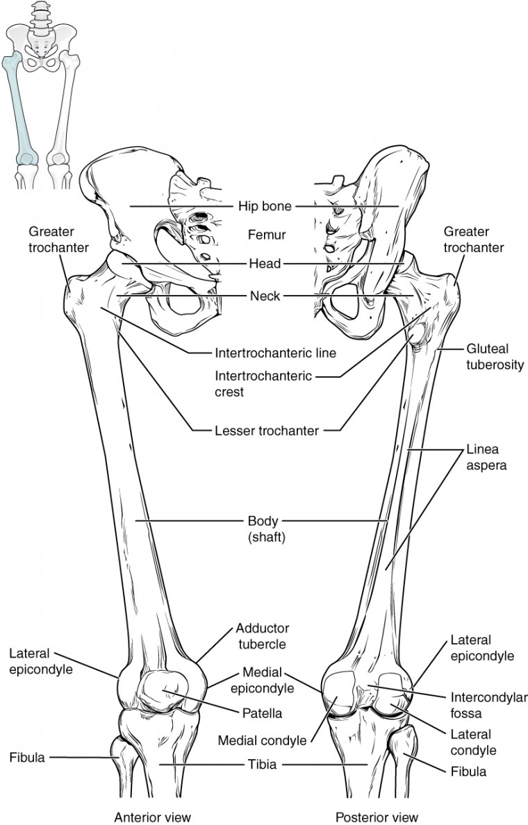
Bones of the Lower Limb | Anatomy and Physiology from s3-us-west-2.amazonaws.com Nov 28, 2016 · anatomy of the larynx. On the left is an anatomy diagram of the internal organs of a female elephant. The larynx, commonly called the voice box or glottis, is the passageway for air between the pharynx above and the trachea below. Click on the image for a larger look at it. It is made up of multiple pieces of tough cartilage, surrounded and held together by fibrous tissues, membranes, and ligaments 3.the largest cartilaginous segment is called the thyroid cartilage, with a prominent bulging known as adam's apple. It is divided into the left and right side by a muscular wall called the septum. The right and left sides of the heart are further divided into two top chambers called the atria (also termed the right and left atrium ), which receive blood and then pump it into the two bottom. Figure 11.32 muscles of the lower leg the muscles of the anterior compartment of the lower leg are generally responsible for dorsiflexion, and the muscles of the posterior compartment of the lower leg are generally responsible for plantar flexion.
Jul 27, 2021 · thoracic wall the first step in understanding thorax anatomy is to find out its boundaries.
Ingested food exits the small intestine and enters the cecum. We'll go over its different openings and functions before exploring the conditions that can affect the diaphragm. The lateral and medial muscles in both compartments invert, evert, and rotate the foot. Jul 30, 2018 · the diaphragm is an important muscle that helps you breathe in and out. The larynx, commonly called the voice box or glottis, is the passageway for air between the pharynx above and the trachea below. Jul 27, 2021 · thoracic wall the first step in understanding thorax anatomy is to find out its boundaries. The upper part of the human nose is quite hard and bony, with the nasal bones forming the nose bridge, while the sides of the upper external nose are supported by the frontal processes and medial plates of the maxilla 12. The right and left sides of the heart are further divided into two top chambers called the atria (also termed the right and left atrium ), which receive blood and then pump it into the two bottom. Jan 21, 2018 · the transverse colon is the lengthy, upper part of the large intestine. On the left is an anatomy diagram of the internal organs of a female elephant. Figure 11.32 muscles of the lower leg the muscles of the anterior compartment of the lower leg are generally responsible for dorsiflexion, and the muscles of the posterior compartment of the lower leg are generally responsible for plantar flexion. As digestion continues, the ingested matter moves up the. Click on the image for a larger look at it.
Share :
Post a Comment
for "Lower Back Organ Anatomy Diagram : Kidney pain vs. Right, Left and Central Abdominal Pain - La vaca cega"


Post a Comment for "Lower Back Organ Anatomy Diagram : Kidney pain vs. Right, Left and Central Abdominal Pain - La vaca cega"